Structure and Composition of Muscle
Objectives:
(1) To provide some insight on the structure of muscle and associated tissues.
(2) To familiarize the student with the nomenclature associated with muscle, connective tissue, adipose tissue, and bone.
(3) To describe the differences among red, intermediate and white muscle fibers.
Reading material: Principles of Meat Science (5th Edition), chapter 2, pages 7 to 52.
Muscle tissue — constitutes the bulk of the carcass of meat animals.
Skeletal muscle — of principal interest to the meat industry. Muscle that is attached directly or indirectly to the skeleton.
Cardiac muscle — muscle of the heart. Differentiated by the presence of intercalated disks.
Smooth muscle — located in arteries and the lymph system as well as the digestive and reproduction systems. No real ordered myofibrils and thus nonstriated appearance.
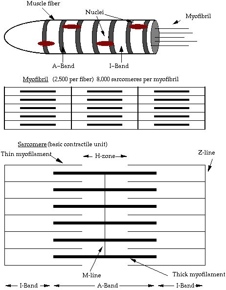
Skeletal muscle fiber
Sarcolemma — membrane surrounding the muscle fiber.
Transverse tubules — portion of the sarcoplasmic reticulum that stores and releases calcium during contraction and relaxation.
Myoneural junction — where motor nerve endings terminate on the sarcolemma.
Motor end plate — structure present at the myoneural junction that forms a small mound on the surface of the muscle fiber
Sarcoplasm — cytoplasm of muscle fibers.
Nuclei — “brain” of the cell. Muscle fibers contain many nuclei.
Myofibrils — long, thin, cylindrical rods, usually 1-2 µm in diameter, that run within and parallel to the long axis of the muscle fiber.
Myofilaments — comprised of thick and thin filaments. The thick are comprised of myosin and the thin are comprised of actin, troponin, and tropomyosin.

Sarcomere — the basic contractile unit of the muscle. Has Z-lines on either end along with A-band and two 1/2 I-bands.
Z-disk ultrastructure — comprised of Z filaments. These are the connecting units between sarcomeres.
Proteins of the myofilament — primarily actin and myosin (65% of total), but also include tropomyosin and troponin within the thin filament, C protein (which surrounds the myosin filaments to form the thick filaments), desmin (which encircles the Z disks and radiate out to connect adjacent myofibrils)
Sarcoplasmic reticulum and T tubules — membranous system of tubules and cisternae (flattened reservoirs for Ca++) that forms closely meshed network around each myofibril.
Mitochondria — “powerhouse of the cell.” Provides the cell with chemical energy.
Lysosomes — small vesicles located in the sarcoplasm that contain a large number of enzymes collectively capable of digesting the cell and its contents. The best known of these are the cathepsins.
Golgi complex — many of these are located in the muscle fiber and serve the same purpose as those in regular cells.
Connective tissue
Extracellular substance — varies from a soft jelly to a tough fibrous mass.
Connective tissue proper — fibrous connective tissue that surrounds muscles, muscle bundles and muscle fibers.
Supportive connective tissues — bone and cartilage.
Ground substance — viscous solution containing soluble glycoproteins (carbohydrate containing proteins) where the extracellular fibers are embedded.
Extracellular fibers — Primarily comprised of collagen, elastin, and reticulin.
Adipose tissue
White versus brown fat — most of adipose tissue in meat animals is white fat. Brown fat is mostly present in animals at birth.
Bone
Diaphysis — long, central shaft of the bone.
Epiphyses — enlargements on the ends of bones.
Periosteum — thin membrane connective tissue covering of bone.
Articular cartilage — present on the ends (joint) of bones. Comprised of hyaline cartilage.
Epiphyseal plate — cartilaginous region separating the diaphysis and epiphysis.
Muscle organization and construction
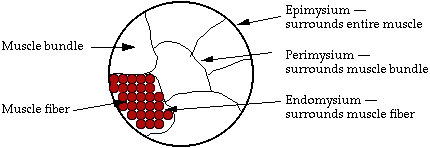
Muscle bundles and associated connective tissues
Endomysium — connective tissue covering of muscle fibers.
Perimysium — connective tissue covering of muscle bundles.
Epimysium — connective tissue covering of entire muscle.

Nerve and vascular supply
Intramuscular fat — deposited within the muscle in loose network of perimysial connective tissue in close proximity to blood vessels.
Intermuscular fat — fat deposited between muscles.
Muscle-tendon junction
Myotendinal — where muscle fibers, bundles and muscles and tendons come together.
Aponeuroses — tendinal attachments of muscles.
Muscle and fiber types
The following chart shows the relationship between the different muscle fiber types:
Characteristics of muscle fibers in domestic meat animals and birds
| Characteristics | Type I | Type IIA | Type IIX(D) | Type IIB |
|---|---|---|---|---|
| Redness | ++++ | +++ | + | + |
| Myoglobin content | ++++ | +++ | + | + |
| Fiber diameter | + | + | +++ | ++++ |
| Contraction speed | + | +++ | +++ | ++++ |
| Fatigue resistance | ++++ | +++ | + | + |
| Contractile action | Tonic | Tonic | Phasic | Phasic |
| Mitochondria number | ++++ | +++ | + | + |
| Mitochondria size | ++++ | +++ | + | + |
| Capillary density | ++++ | +++ | + | + |
| Oxidative metabolism | ++++ | ++++ | + | + |
| Glycolytic metabolism | + | + | +++ | ++++ |
| Lipid content | ++++ | +++ | + | + |
| Glycogen content | + | + | ++++ | ++++ |
| Z disk width | ++++ | +++ | + | + |
Adapted from Table 2.1, Principles of Meat Science (5th Ed.), page 45.
Chemical composition of the animal body
Water — Fluid medium of the body
Proteins — Structure and metabolic reactions in body
Lipids — Sources of energy, cell membrane structure and function, and metabolic functions (vitamins and hormones)
Carbohydrates — low level in body, mainly glycogen in muscle and liver
Review of Material — What the student should know:
(1) The terminology associated with describing muscle and muscle components.
(2) The various components of the muscle and structures closely associated with it.
(3) The specific and general components of muscle and associated structures.
(4) The differences in structure and metabolism in the different muscle fiber types.
Links to related sites on the Internet
Growth and Development of Meat Animals – Howard Swatland, author
J.W. Savell, revised January, 2016
